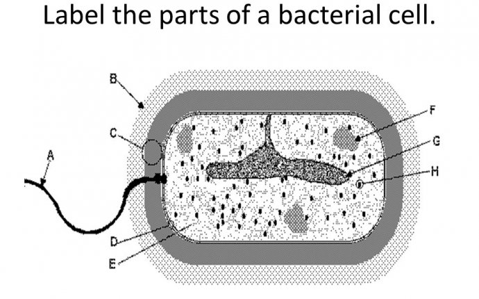
Parts of a Bacteria cell
Back in 1884 a guy named Gram developed a staining technique to visualise bacterial samples (now called a Gram stain). It was really important because, as the story goes, pneumonia was a big problem at the time and there were three causes; unknown (later identified as viral pneumonia) and two types of bacterial pneumonia caused by either Streptococcus pneumoniae or Klebsiella pneumoniae. Importantly pneumonia caused by Streptococcus is more contagious and develops faster than pneumonia caused by Klebsiella, which tends to only affect the immuno-compromised. Gram’s stain, which was fast and definitive, allowed for the three different types of pneumonia patient to be grouped together, reducing spread and therefore preventing disease.
So how did Gram’s stain work? Because of the peptidoglycan layer. The thickened peptidoglycan layer in Gram positive cells allows them to retain the stain (hence remaining ‘stain positive’ or ‘Gram positive) where as the thin layer seen in Gram negative cells cannot prevent the stain from leeching out (hence stain and Gram negative). Of course Gram himself didn’t know this but his stain was a success and it was 1884 so give him a break.
Peptidoglycan is also vitally important for the way antibiotics work. The role of a bacterial cell wall is defensive. The wall is there for the same reason our skin is on us, to keep the insides in and the outsides out and it does this by physically limiting the size and shape of the cell. In the microbial world one of the most important forces changing cell size and shape is, believe it or not, water.
A bacterial cell is a little salty bubble generally existing in a less salty environment. The problem lies in that the less salty environment wants to even out all the salt concentrations so water would rush into the cell to dilute its saltiness until it matches that of the environment, or until it bursts and kills the cell. This process is given the name osmosis. The role of peptidoglycan is to act as a physical barrier to the cell taking on to much water and killing itself. Its like trying to inflate a balloon inside a small box, once a certain amount of air goes in the box pushes back on the expanding balloon and no more air can be pushed into the balloon.
But suppose we could break this peptidoglycan wall, that would result in the bacterium losing this protective layer and becoming vulnerable to osmosis causing the cell pop. Wouldn’t that be a great antibiotic?
Turns out it is a great antibiotic, penicillin. Penicillin works by inhibiting the repair of the peptidoglycan layer, therefore damage compounds and the peptidoglycan is compromised causing it to become susceptible to osmotic lysis.
This also explains why penicillin and its derivative are more effective against Gram positive cells. With its peptidoglycan layer hidden beneath an outer lipid membrane it is harder for the penicillin to reach the peptidoglycan where it has activity whereas Gram positive cell walls leave the peptidoglycan exposed.
Penicillin is so good at killing bacteria that bacteria have had to evolve a way around it. They do this in two ways, they either destroy the penicillin itself or they change the target of penicillin to something penicillin can’t recognise. Either way our use of penicillin, and our exploitation of this peptidoglycan wall triggered an arms race with the microbial world so that they could protect the precious peptidoglycan.
I mentioned at the top that S. aureus knows what is grandparent looked like and that this was related to peptidoglycan and this comes back to how this bacteria determines how it will divide.
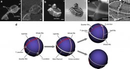
A recent paper in Nature Communications by Prof. Simon Foster’s group (Turner et al., 2010, see below) has shown that the Golden Staph has detectable ridges in its peptidoglycan structure, a kind of pie crust that can be found in a very specific pattern. They found that one ridge was equatorial (whole rib), a second ridge bisected only one hemisphere (half rib) and a third ridge perpendicularly bisected one half of the previously bisected hemisphere (quarter rib).

Its been known for some time that Staphylococcus forms in bunches, in fact it name comes from the Greek word for grapes, and even more recently it has been observed that staphylococcal cell division takes place in a very specific order. The first division is within the -axis, the second within the y-axis then the third in the z-axis before repeating itself. Each cell division takes place within a new plane and at right angles to the last cell division.
What Prof. Foster and his group have shown is that the pie-crusts or peptidoglycan ribs mark the site of peptidoglycan synthesis during Staphylococcal cell division and because of the way each cell divides it retains the information of the two previous divisions, its parental and grand-parental divisions! Furthermore, this observation indicates this process is not random and so probably driven by the peptidoglycan itself.
Peptidoglycan is a wonderful substance. Without it bacteria would be vulnerable to death by water, we wouldn’t be able to quickly, easily or cheaply tell them apart and we would be without penicillin, possibly the second greatest biomedical innovation after vaccines. Now it seems that peptidoglycan can control the site of cell division, in S. aureus anyway, indicating there might be more to discover about this bacterial wonderwall.
References:
Turner, R., Ratcliffe, E., Wheeler, R., Golestanian, R., Hobbs, J., & Foster, S. (2010). Peptidoglycan architecture can specify division planes in Staphylococcus aureus Nature Communications, 1 (3), 1-9 DOI: 10.1038/ncomms1025
van Heijenoort J (2001). Formation of the glycan chains in the synthesis of bacterial peptidoglycan.


